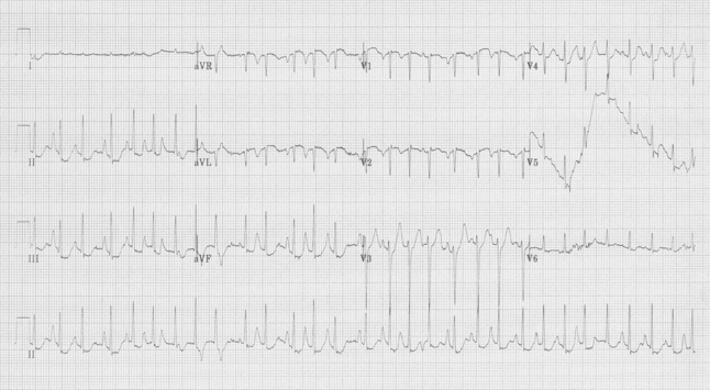
 64-year-old woman with COPD complains of nausea.
Interpretation & Explanation
An irregularly irregular tachycardia at 180/minute, this rhythm could be mistaken for atrial fibrillation, but notice the large upright waves between QRS complexes – these are not T waves, but are P waves of varying morphology. They are so large in a lead II rhythm strip because this woman with COPD has R atrial enlargement and these are resulting “P pulmonale”. This rhythm is multifocal atrial tachycardia (MAT).

MAT – rapid, irregularly irregular rhythm with P waves of varying morphology
MAT can be caused by several conditions including: electrolyte abnormalities, exacerbation of respiratory disease, drug toxicity (theophylline toxicity), and almost any severe illness – it is said that MAT is one of the common arrhythmias noted in an ICU population.
This patient had recently been discharged home after an admission for exacerbation of COPD, but had no respiratory complaints on this presentation. Her theophylline level was 40μg/mL (>2x normal), and a review of her discharge medications found an incorrectly written prescription for twice the usual daily dose of theophylline.