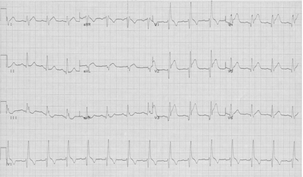
65-year-old man with substernal chest pain.
This gentleman had chest pain and associated symptoms suggestive of an acute coronary syndrome. His EKG reveals STEMI in leads V2 – V5. This is an important pattern to recognize. A RBBB does not perturb the depolarization very much (because the RBBB supplies the much smaller side of the heart), certainly much less than LBBB. Because the depolarization is less altered it is expected that ST-segment changes of acute ischemia or infarct will be recognizable on the EKG.
If there are any questions about the ST-segment elevation in V2-3 remember that the horizontal tracings on the 12-lead are simultaneously recorded, and that complexes line up vertically. The beginning and the end of the QRS is clear in V1, and if you follow the end of this QRS down through V2-3, the ST-segment in these complexes becomes clear – it is not just a wide QRS in these leads!
Another RBBB with acute anterior STEMI – perhaps more clearly seen

