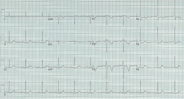
56-year-old woman with prior chest pain six hours ago.
Patient was pain free at the time of this EKG.
The most striking finding on this EKG is the deep T-wave inversions in V2-3 and the biphasic T wave in V4. She was pain-free at presentation and an initial troponin was negative. Three hours later the repeat EKG was unchanged and a second troponin was minimally elevated at 0.12. She was heparinized and admitted to telemetry. She went to cardiac cath the next morning – that revealed 90% proximal LAD stenosis that was successfully stented.
Since these anterior T-wave inversions appeared in the pain-free interval following her chest pain episode, I believe they represent Wellens’ syndrome. Wellens’ may present with biphasic T-waves in the anterior leads or with deeply inverted T-waves in the same leads (original reports actually claimed deep T-wave inversion were the more common finding). We can add this form of Wellens’ to the differential list of deeply inverted T waves (remember: spasm, Takotsubo, CNS event, and rarely PE).
Wellens noted that unstable angina with this EKG pattern frequently (75% of his patient series) developed an extensive anterior MI with days to weeks of presentation. This finding correlates with angiography in the modern era that typically demonstrates a critical lesion in the proximal LAD.
Rhinehardt J, Brady WJ, Perron AD, et al. Electrocardiographic manifestations of Wellens’ Syndrome. Am J Emerg Med 20:638-643, 2002.
Tandy TK, Bottomy DP, Lewis JG. Wellens’ Syndrome. Ann Emerg Med 33:347-351, 1999.