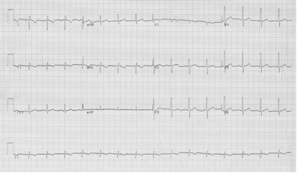
What is the diagnosis associated with this EKG?
Answer & Explanation
This EKG has several unusual findings – 1) a significant right axis deviation, 2) upright QRS in lead aVR, and 3) inappropriate R-wave progression in the chest leads. Remember that lead aVR is oriented opposite to other limb leads, and always has a negative P, QRS, and T wave in a normal EKG. The appearance of a negative complex in I and upright complexes in aVR can be seen with switched limb leads…

but notice that the R-wave progression of the chest leads is normal when just limb leads are switched.
This challenging EKG is from a patient with dextrocardia. It is advisable to place the chest leads in the right-sided array to get an improved diagnosis of ischemia and infarct patterns.