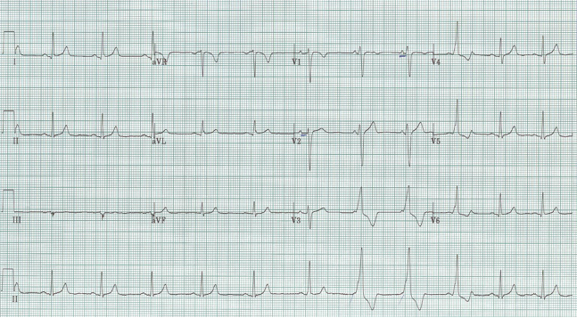
What is the rhythm change seen on this tracing?
Baseline is sinus rhythm at 68/minute
Normal axis and intervals
Essentially normal EKG with several strange QRS complexes…

First, if you march the P waves through, they remain constant (note the pen marks of some helpful person). The 4th and 5th beats in this rhythm strip are wide-complex and dissociated from the P waves. This rhythm is sometimes called “slow v tach” to indicate the independent (ectopic) ventricular source. This is accelerated idioventricular rhythm (AIVR).
Another helpful feature of this ventricular rhythm are fusion beats as the rhythm phases in and out with sinus rhythm, often at a similar heart rate. In this rhythm strip, the 3rd and 6th beats are fusion beats – that is, a combination of the sinus beat and the ventricular beat.
AIVR is well known as a repolarization arrhythmia (especially after TPA or interventional cath). It can also occur in essentially normal patients for no apparent good reason, as was the case in this otherwise healthy young woman.