50-year-old woman, a chronic alcoholic, with three days of nausea, vomiting and diarrhea, presents after syncope.
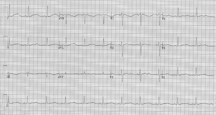
EKG interpretation:
Sinus at 70/minute
Axis normal (10°)
PR and QRS intervals normal, QT is prolonged in many leads (eg aVL)
No chamber enlargement
A U wave is present after the T wave in several leads (leads V2-3)
The important features to recognize in this EKG is the appearance of a U wave and the diminution of the T-waves, leaving the appearance of a prolonged QT in several leads. The differential of a prolonged QT interval, besides offending medications, are hypokalemia (seen with increasing U waves and diminishing T waves), hypomagnesemia, and hypocalcemia. Alcoholics are already prone to hypomagnesemia, and the addition of several days of gastroenteritis may quickly deplete other electrolytes and cations.
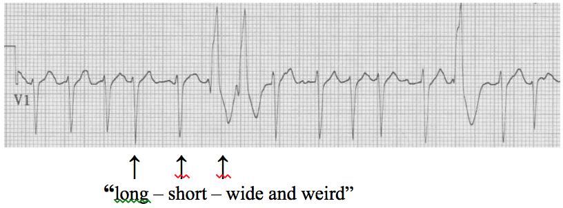
While the patient was being monitored during the initial stages of her evaluation, she suddenly became unresponsive and the following rhythm was captured on the monitor:

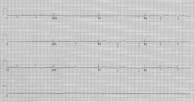
She regained consciousness with sinus rhythm on the monitor and stable VS without intervention. Soon thereafter labs resulted:
K – 2.3 mEq/L (3.6 – 5.2)
Mg – 0.6 mEq/L (1.3 – 1.9)
iCa – 0.96 mmol/L (1.1 – 1.3)
Remember that alcoholics who become sick are at risk for prolonged QT, torsade de pointe, and syncope or sudden death!


