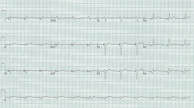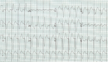
Does this ventricular paced complex look different from usual?
Answer & Explanation

Typical ventricular paced complexes – since the pacing lead is inserted into the right ventricle the current of depolarization proceeds from right to left ventricle effectively producing a wide QRS with LBBB morphology.
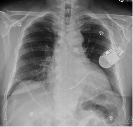
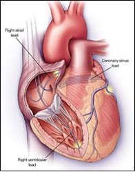
Today’s EKG is 100% ventricular paced with a fairly narrow and normal appearing QRS complex. This patient has a biventricular pacemaker with one lead in the RV (also his ICD lead), and a second wire into the coronary sinus (thinner lead extending further to the left heart wall). This pacer produces simultaneous biventricular activation and is often used now in patients with significant heart failure.

