This is an excellent example of EKG changes of COPD.
Findings suggestive of COPD include (but you don’t need to have all):
- P waves >0.25 mV in II, III, or aVF (P “pulmonale”)
- Lead I sign – isoelectric P wave, QRS amplitude <0.15 mV, and T wave <0.05mV in lead I
- QRS amplitude in all limb leads <0.5 mV
- QRS axis > 90° (right axis deviation)
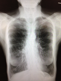
Just in case you’re still not a believer, here’s her CXR…

Note the impressive changes of emphysema: flat and depressed diaphragms and the “vertical” heart.