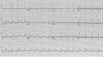
First the rhythm – sinus bradycardia at 36/minute
Second – the inferior leads keep catching my eye…no obvious ST-segment elevation, but the T waves look a bit prominent (hyperacute ?), and then there is the T wave inversion in aVL (remember, reciprocal changes in aVL may be the earliest sign of an inferior MI).
Of course, the correct response would be to repeat the EKG. This was done just 10 minutes later:

Now the inferior STEMI is evident! Note the rhythm change – now sinus at 100/minute, and with more prominent P waves. Not having been there, I can only guess – the original rate was likely due to excess vagal tone (common with inferior MI’s – think pale, diaphoretic, nauseous, bradycardic). The subsequent tachycardia might be due to nitroglycerin administration, or just less vagal tone, or maybe they received atropine.
Keep repeating those EKGs when rhythm or rate changes; you could be looking at an early MI
